Nanoparticles used for drug delivery to a particular part of the body are often broken down prematurely by the liver. Jeroen Bussmann, the chemical biologist at Leiden University, has reported a new approach for preventing this from happening in the ACS Nano journal.
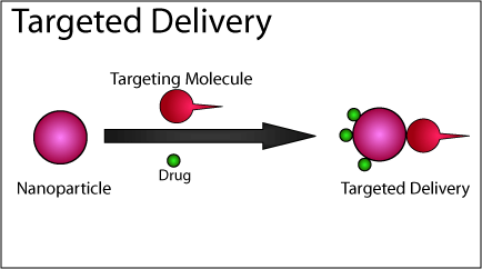
In nanotherapy, micro-nanometer-sized particles are used to transport drugs to specific locations in the body; for instance, to kill tumor cells. They have far fewer side-effects compared to conventional chemotherapy. However, a recurrent issue in developing nanotherapy is that nanoparticles are often broken down prematurely in the liver. As a result, the chance for the nanoparticles to reach their intended locations is rare. Until now, scientists believed that this process was the work of Kupffer cells, the so-called clean-up cells in the liver.
Cells from Blood Vessel Walls
In collaborative research work conducted with the Hubrecht Institute and the University of Basle, Jeroen Bussmann reported that cells in the liver’s blood vessel walls (endothelial cells) usually play a key role in this process. The nanoparticles are recognized and eliminated by the proteins on the surface of these cells. If these proteins are blocked, then the nanoparticles will no longer be broken down by the endothelial cells and therefore, they can remain in the blood for a longer period. This is an important step for delivering drugs to their intended destinations in the body.
Tracking Nanoparticles
In this study, zebrafish larvae were used by Bussmann. “The advantage of using these larvae is that they are transparent, so we can follow the nanoparticles as they move through the blood vessels using a microscope,” explains Bussmann. He blocked the endothelial cells by providing the zebrafish larvae a special polymer – which was a long, interlinked molecule. “When this polymer binds to the proteins on the endothelial cells, they no longer recognize the nanoparticles,” he explains.
In the liver, the other clean-up cells (Kupffer cells) predominantly identify particles with a size of more than 100 nm. The idea was that by using the combination of smaller nanoparticles and the special polymer, no more cells would be present in the liver to eliminate the nanoparticles. This approach worked: Nanoparticles administered in this manner are not broken down and remain unaffected in the bloodstream.
Blood Vessel Cells Swallow Nanoparticles
The moment Bussmann could be confident the nanoparticles had been actually ingested by the endothelial cells, was when the fish larvae were administered with nanoparticles containing a toxic substance, which acts only inside the cells but not outside of them. Hence, when only the endothelial cells were killed, Bussmann concluded that their cause of death was due to the ingestion of nanoparticles.
With the zebrafish larvae, Bussmann was also able to find exactly which protein in the endothelial cells attaches to the nanoparticles – namely Stabilin-2. Moreover, the removal of the gene for Stabilin-2 led to the much lower breakdown of the nanoparticles. At present, Bussmann is planning to create a molecule that attaches specifically to the Stabilin-2 protein. As a result, the breakdown function of the cells can be inhibited highly specifically, while at the same time, the liver does not lose part of its natural function.
Delivering Medicines to Cells
Furthermore, Bussmann wants to investigate how precisely the protein attaches to the nanoparticles and the subsequent ingestion of the nanoparticles by the endothelial cells. “We want to understand every step in the process so that we can ultimately produce nanoparticles that can deliver medicines not only to the liver but to every type of cell in the body.”
Source : https://www.azonano.com/news.aspx?newsID=36049